Ultrasound in Urology
What types of ultrasound are there in urology?
Depending on the organ to be examined and the medical question, different ultrasound techniques are used:
- Abdominal ultrasound: The standard method for examining the kidneys, ureters, and bladder through the abdominal wall.
- Transrectal ultrasound (TRUS): A special examination technique for detailed assessment of the prostate via the rectum.
- Doppler and color duplex sonography: This method measures blood flow in vessels and is particularly used for vascular constrictions or testicular torsions.
- Scrotal ultrasound: Examination of the testes and epididymis to clarify pain, swelling, or tumors.
Applications of Urological Ultrasound
Ultrasound is used in urology for various diagnostic purposes:
- Kidneys and urinary tract: Examination for kidney stones, tumors, cysts, or abnormalities in the ureters.
- Bladder: Detection of urinary outflow disorders, bladder stones, or tumors.
- Prostate: Examination for an enlarged prostate (benign prostatic hyperplasia), prostate cancer, or inflammations.
- Testicles and penis: Diagnosis of testicular torsion, epididymitis, tumors, or vascular changes.
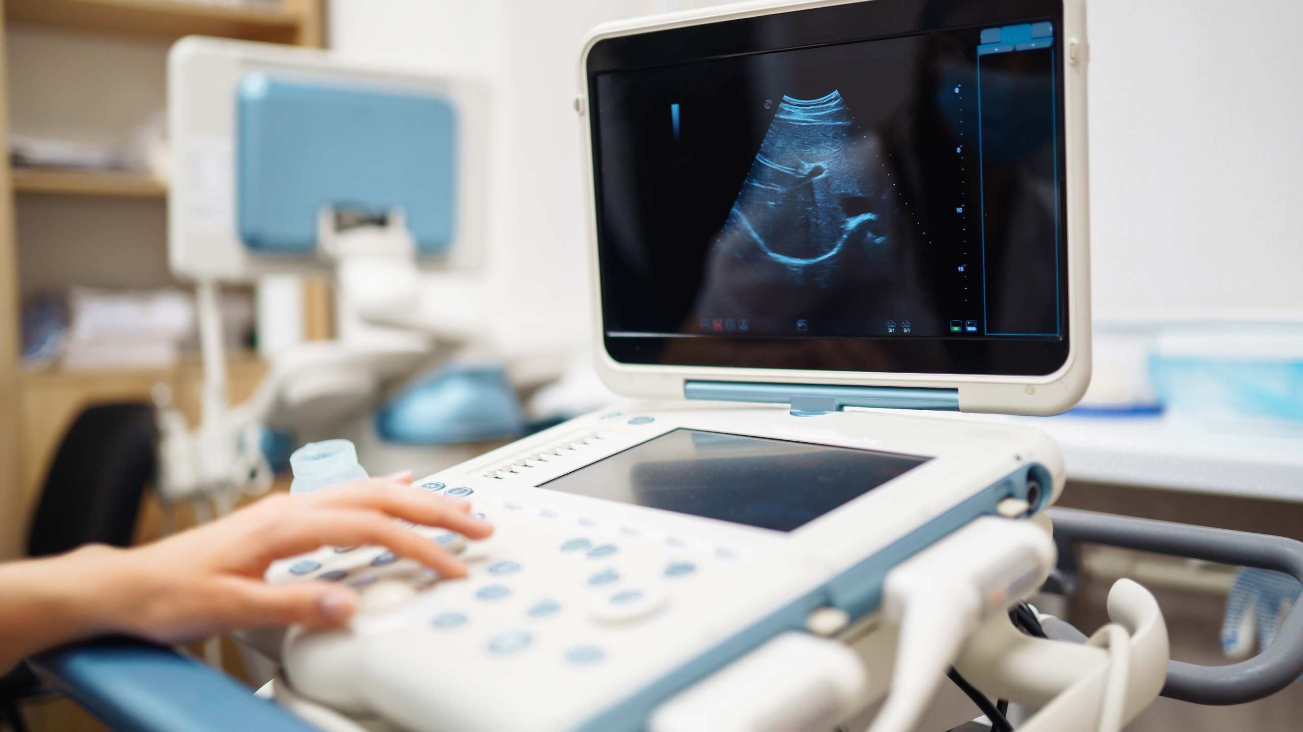
How does a urological ultrasound examination proceed?
The process of an ultrasound examination is simple and comfortable for the patient. Depending on the organ to be examined, the sonography is performed either through the abdominal wall or via a body opening:
- The patient lies relaxed on an examination couch.
- The doctor applies a special contact gel to the skin to ensure better conductivity of the sound waves.
- With the ultrasound probe, the doctor examines the organs to be checked. Real-time images are produced on a monitor.
- In transrectal ultrasound (TRUS), a thin ultrasound probe coated with gel is carefully inserted into the rectum.
- The examination usually does not last longer than 5 minutes.
What are the advantages of ultrasound in urology?
- Non-invasive and painless: No needles, no incisions.
- Radiation-free: Unlike X-rays or CT scans, there is no harmful radiation exposure.
- Real-time imaging: Dynamic processes such as bladder emptying or blood flow can be observed.
- Fast and cost-effective diagnosis: An ultrasound device is available in every urological practice and provides immediate results.
- Early detection of diseases: Tumors, inflammations, or malformations can be detected at early stages.
When is further diagnostics necessary?
If the ultrasound examination shows abnormalities, further imaging procedures may be necessary:
- Computed tomography (CT) or magnetic resonance imaging (MRI) for a more detailed assessment of tumors or complex diseases.
- Urodynamic studies if functional disorders of the bladder or urethra are suspected.
Related News

Cavernous Body Ultrasound
The corpus cavernosum ultrasound, also called penisonography, is an imaging procedure for assessing the structure and blood flow of the corpus…

Urodynamics
Urodynamics includes diagnostic procedures for assessing the storage and voiding function of the bladder and urethra. It helps identify functional…

Shockwave Therapy for Erectile Problems - ESWT
Shockwave therapy (ESWT) has established itself as an innovative and effective method for treating erectile dysfunction. This non-invasive procedure…